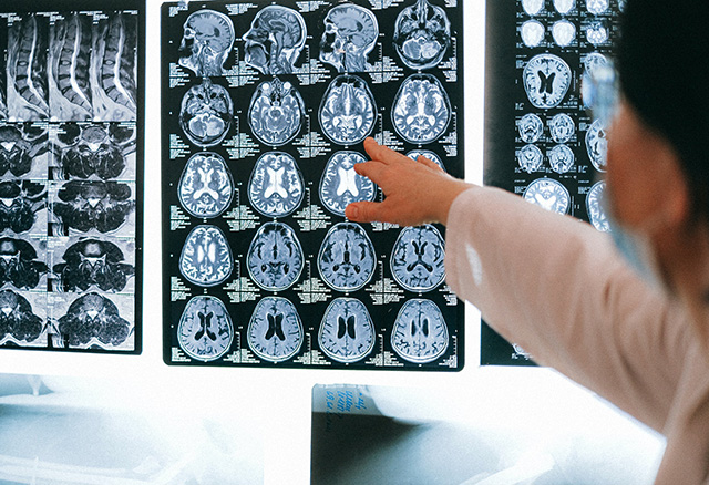
Using magnetic resonance imaging (MRI) is just one way of looking at body composition. MRI is a noninvasive, painless medical imaging technique used commonly in radiology. MRI provides detailed images of the body in any plane, and is used to visualize the structure and function of the body for the diagnosis and treatment of human conditions. Due to the detailed images that can be provided using this technique, it is possible to get accurate measurements of the composition of body tissue.
purpose: MRI, that is used to diagnose and treat medical conditions. In terms of body composition, the high-quality images can be processed to differentiate and measure the amounts of fat and lean body tissue and their distribution.
equipment required: MRI Scanner
 MRI scan results
 method: MR imaging uses a powerful magnetic field, radio frequency pulses and a computer to 'excite' water and fat molecules in the body, producing detailed pictures of organs, soft tissues, bone and virtually all other internal body structures.
procedure: A person lies within the magnet as a computer scans the body, which can take about 30 minutes. High-quality images show the amount of fat and where it is distributed.
advantages: this is a noninvasive method for body composition analysis
disadvantages: The use of MRI is limited due to the high cost of equipment and analysis.
other comments: this technique does not use ionizing radiation (unlike CT scans, x-rays), so is very safe.
Similar Tests
Related Pages
- Other body composition tests
- About measuring body composition


 Current Events